
Hori’s Naevus
Hori’s naevus sits deeper within the skin, which is why the right treatment starts with understanding it
Introduction
What Is Hori's Naevus?
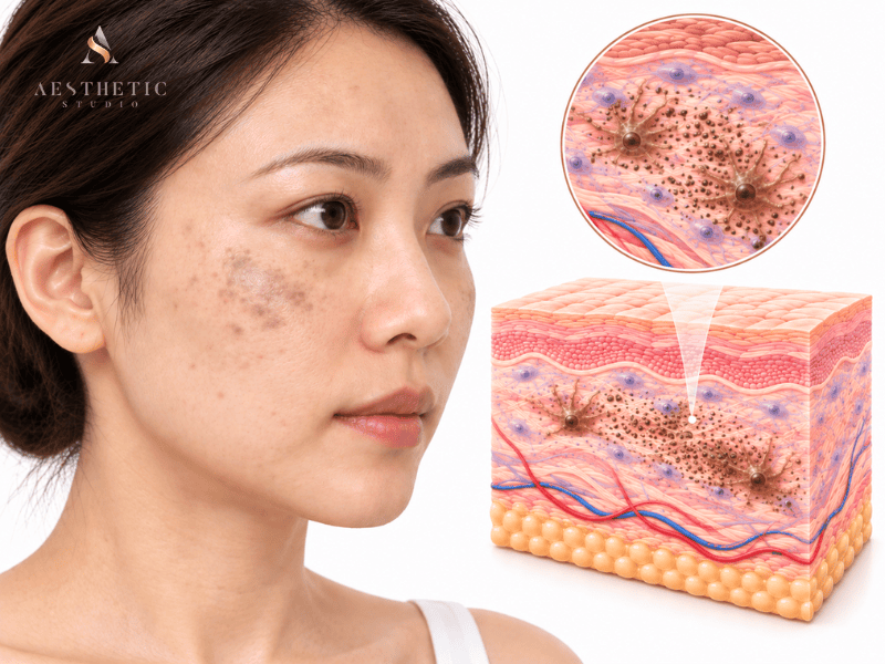
Hori’s naevus is a type of acquired dermal pigmentation that typically appears as small blue-grey, grey-brown or slate-coloured spots across both cheeks. Also known as acquired bilateral naevus of Ota-like macules (ABNOM), it commonly develops in adulthood and is seen more often in Asian skin.
Because the pigment sits deeper within the dermis, Hori’s naevus can be more persistent than superficial pigmentation such as freckles or sun spots. It can also look similar to melasma, particularly when pigmentation appears across the cheekbones — and some people may have more than one type of pigmentation at the same time.
Recognising Hori’s naevus, and understanding how it differs from other types of pigmentation, is important because where the pigment sits can influence how it is treated.
Key Signs
What Does Hori's Naevus Look Like?
Hori’s naevus has a characteristic pattern, although it can sometimes resemble other forms of facial pigmentation. Looking at the colour, location and pattern of the spots can provide useful clues.
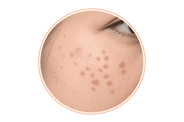
Grey, blue-grey or brown-grey spots
Hori’s naevus often appears as grey, blue-grey or grey-brown pigmentation, rather than a straightforward brown. The exact shade can vary depending on how deeply the pigment sits within the skin.
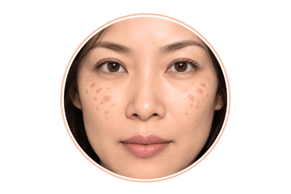
Usually appear on both cheeks
A typical pattern involves pigmentation on both sides of the face, particularly across the upper cheeks and cheekbones. The distribution may be similar on both sides without being perfectly symmetrical.
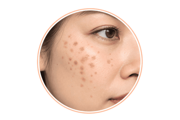
Small spots that may cluster together
Rather than one uniform patch, Hori’s naevus often appears as multiple small, speckled spots or macules. As these cluster together, they can resemble a broader area of facial pigmentation.
Causes
What Causes Hori's Naevus?
There is no single confirmed cause of Hori’s naevus. Clinical research suggests that it may involve pigment-producing cells called melanocytes within the dermis becoming active, with several biological and environmental factors proposed to influence this process.¹,²
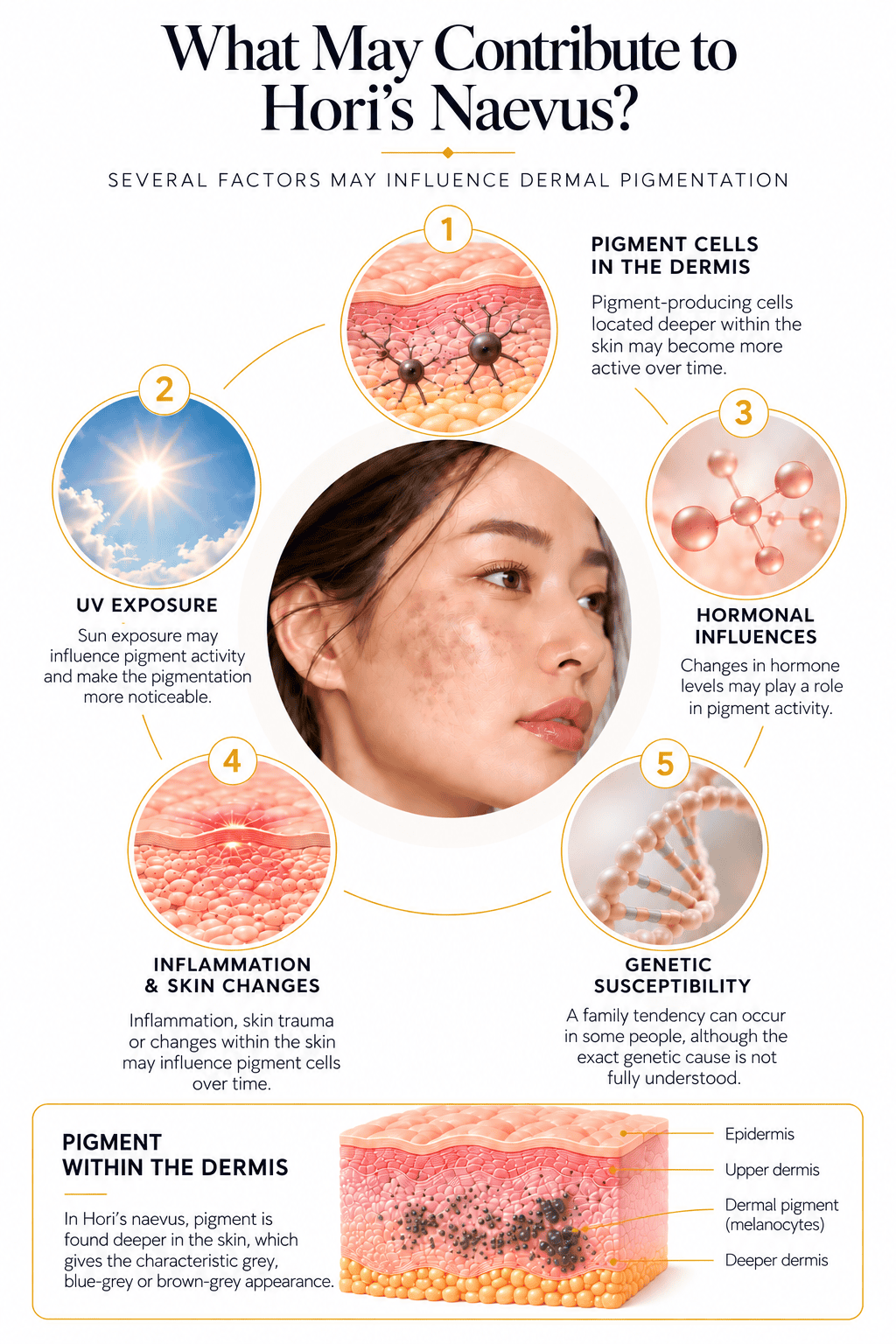
Deeper Pigment Cells Becoming Active
Hori’s naevus involves pigment-producing cells located deeper within the skin. When these cells become more active, they produce melanin within the dermis, which can gradually appear as grey, blue-grey or grey-brown pigmentation on the cheeks.
UV and Sun Exposure
Repeated UV exposure may stimulate pigment activity and make Hori’s naevus more noticeable over time. This is one reason daily sun protection remains important, even though sun exposure itself is not considered the sole cause of the condition.
Hormonal Changes
Hormonal changes may influence pigment activity, which may help explain why Hori’s naevus can first become noticeable during adulthood. For some people, the pigmentation appears gradually and becomes more apparent over time rather than developing suddenly.
Inflammation and Skin Changes
Inflammation or changes within the skin may also influence pigment-producing cells. This is particularly relevant when different types of pigmentation occur together, as what appears to be one area of pigmentation may have more than one underlying component.
Genetic Susceptibility
Some people may simply be more predisposed to developing Hori’s naevus than others. A family tendency can occur, although having a relative with Hori’s naevus does not necessarily mean that you will develop it too.
Treatments
Treatment Options for Hori's Naevus
Because Hori’s naevus involves pigment within the deeper dermal layer of the skin, treatment requires a different approach from more superficial forms of pigmentation. Picosecond and Q-switched lasers are among the pigment-specific technologies used to target this deeper melanin.
The appropriate treatment is not determined by the laser alone. Our doctors assess the colour, depth and distribution of the pigmentation, together with the patient’s skin type and treatment history, before selecting the laser, wavelength and treatment parameters.
This is especially important in Asian skin, where careful treatment planning can help minimise unnecessary inflammation and the risk of post-inflammatory hyperpigmentation (PIH).
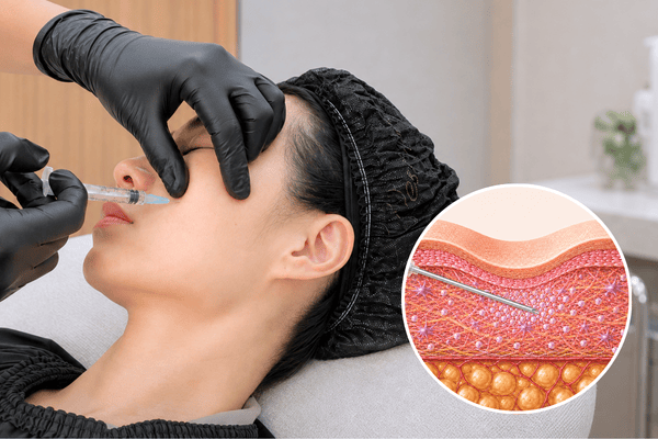
Picosecond Laser
Picosecond lasers deliver energy in ultra-short picosecond pulses, producing a predominantly photoacoustic or photomechanical effect that breaks pigment into smaller particles.
For Hori’s naevus, selected wavelengths can penetrate into the dermis to target the deeper melanocytes and melanin responsible for its characteristic grey, blue-grey or grey-brown appearance. Picosecond lasers have been specifically studied for the treatment of Hori’s naevus and other forms of dermal pigmentation.
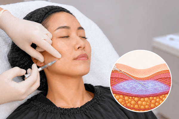
Q-Switched Laser
Q-switched lasers deliver energy in nanosecond pulses that are selectively absorbed by melanin. They have a long-established role in the treatment of dermal pigmentation, including Hori’s naevus.
Different wavelengths penetrate the skin to different depths. For Hori’s naevus, the doctor can select an appropriate wavelength and treatment parameters according to the depth, colour and distribution of the dermal pigment. Q-switched Nd:YAG laser treatment for Hori’s naevus has also been studied specifically in Asian patients.
FAQ
Frequently Asked Questions About Hori's Naevus
How do I know if I have Hori’s naevus?
You may suspect Hori’s naevus if you notice small grey, blue-grey or grey-brown spots appearing on both cheeks, particularly if they developed gradually during adulthood.
However, colour alone is not enough to make the diagnosis. Melasma, post-inflammatory hyperpigmentation and other forms of facial pigmentation can sometimes overlap or look similar.
If you have had persistent cheek pigmentation and are unsure what it is, an assessment with our doctors can help determine the pattern and likely depth of the pigment before deciding how it should be treated.
Is Hori’s naevus the same as melasma?
No. Hori’s naevus and melasma are different pigment conditions, although they can look surprisingly similar when they appear across the cheeks.
Hori’s naevus typically forms multiple discrete grey, blue-grey or grey-brown spots caused by pigment within the dermis. Melasma more commonly appears as broader patches of brown pigmentation and has a different biological behaviour.
The difficulty is that some patients can have both Hori’s naevus and melasma at the same time. This is one reason pigmentation that has been treated as “melasma” may not behave as expected.
Will Hori’s naevus disappear on its own?
Hori’s naevus is a persistent form of dermal pigmentation and typically does not fade completely on its own. In some people, the pigmentation may gradually become more noticeable over time.
If you are noticing persistent grey, blue-grey or grey-brown pigmentation across the cheeks, an assessment can help determine whether it is Hori’s naevus or another type of pigmentation such as melasma.
Once the pigment has been identified, our doctors can advise whether treatment is suitable and which pigment-targeting approach may be appropriate for your skin.
Can creams or skincare remove Hori’s naevus?
Skincare can play an important role in sun protection, reducing unnecessary inflammation and managing other pigmentation that may coexist with Hori’s naevus, but Hori’s naevus itself involves melanocytes deeper within the dermis.
For that reason, topical skincare should not be expected to work in the same way as treatment that directly targets dermal pigment.
This distinction becomes especially important when someone has been using brightening products for what they assumed was melasma or superficial pigmentation without seeing the improvement they expected. Persistent grey-brown cheek pigmentation may warrant reassessment of the diagnosis.
Is picosecond or Q-switched laser better for Hori’s naevus?
There is currently no good evidence that one is universally better for every patient with Hori’s naevus.
Picosecond lasers deliver energy in ultra-short picosecond pulses and produce a strong photomechanical effect that fragments pigment. Q-switched lasers use nanosecond pulses and have a much longer clinical history in the treatment of dermal pigmentation.
A 2026 systematic review found that both technologies can achieve pigment clearance, but it did not establish a statistically significant difference in clearance between the two categories. Importantly, the researchers rated the certainty of the evidence as very low, so this should not be interpreted as proof that the lasers are equivalent.³
Rather than choosing a treatment simply because one laser is newer, the more useful question is which wavelength and treatment approach are appropriate for the pigment and skin being treated.
How many laser sessions are needed for Hori’s naevus?
The number of treatments needed for Hori’s naevus varies from person to person. The best place to start is with an assessment, so our doctors can examine the pigmentation, confirm whether it is Hori’s naevus and determine the appropriate treatment approach.
From there, we can give you a more personalised idea of what to expect and adjust the treatment plan according to how your pigmentation responds.
Medical References
- Park JM, Tsao H, Tsao S. Acquired bilateral nevus of Ota-like macules (Hori nevus): Etiologic and therapeutic considerations. Journal of the American Academy of Dermatology. 2009;61(1):88–93. View study on PubMed
- Ee HL, Wong HC, Goh CL, Ang P. Characteristics of Hori naevus: a prospective analysis. British Journal of Dermatology. 2006;154(1):50–53. View published study
- Li F, Wang Y, Shen Z. Efficacy of Q-Switched and picosecond lasers for acquired bilateral nevus of Ota-like macules: a systematic review and exploratory dose-response meta-analysis. Lasers in Medical Science. 2026;41(1). doi:10.1007/s10103-026-04982-6. View published guidance